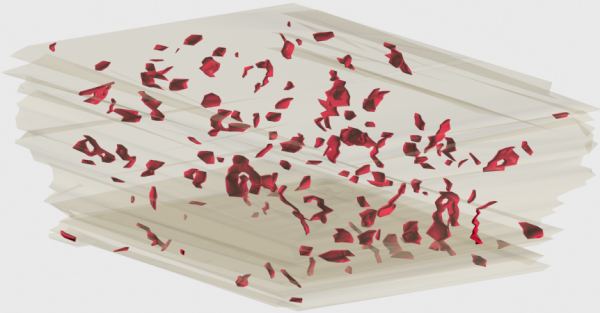
Synapses in stratum radiatum of hippocampal area CA1 are distributed throughout the neuropil. Where an unmyelinated axonal bouton and a dendrite come into to contact there is the potential for a synapse. But only a few of these contacts are synaptic. Figure 1 shows the distribution of synapses within a volume of neuropil from stratum radiatum of the adult rat. There are 161 synapses in this 64 cubic micron volume. These are all chemical synapses, 94% of which are Type 1 (excitatory) synapses and 6% Type 2 (inhibitory) synapses.

Figure 1: Synapses in a Volume of Neuropil
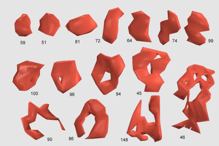
This image was created by reconstructing the synapses in 3D from serially-sectioned tissue that was photographed on the electron microscope. As can be seen in the figure, synapses take on a variety of shapes. This can be more readily seen by viewing a sample of these synapses placed en face (Figure 2).

Figure 2: Variety of Synapse Morphologies
Most synapses cover a small area and have a compact, roughly convex shape, such as numbers 51, 59, and 81, above. These are referred to as macular synapses. Larger synapses are often exhibit "holes" in the middle. These holes are regions of cell membrane devoid of the specializations characteristic of the synapse, e.g. postsynaptic density, synaptic cleft, presynaptic active zone, etc. Synapses with holes, such as numbers 45, 46, 86, 90, 94, 96, and 100, are referred to as perforated synapses. Of the 161 synapses in this volume of neuropil, 148 are macular, while the remaining 13 are perforated.
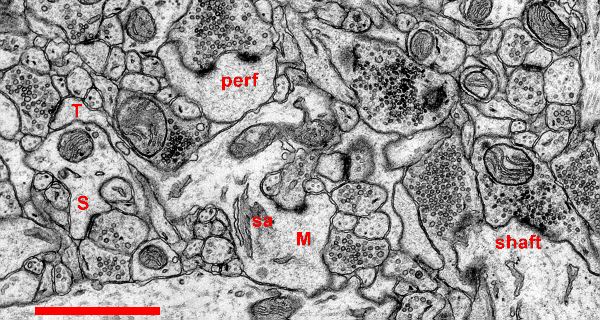
The difference between macular and perforated synapses can be seen in electron micrographs in which the postsynaptic densities have been stained (Figure 3).

Figure 3. Synapses in Hippocampal Area CA1 of the Rat. Scale: 1 micron
Most synapses in stratum radiatum (>90%) occur on spines. As shown in Figure 3, spines come in a variety of shapes. A thin spine (T) has a small head with a macular postsynaptic density. The length of the spine neck is much greater than its diameter. The mushroom spine (M) has a large head, typically greater than 0.6 microns in diameter. An elaboration of the endoplasmic reticulum, called a spine apparatus (sa) is often visible within the neck of a mushroom spine. Mushroom spines also tend to have perforated (perf) postsynaptic densities on the spine head. The stubby spine (S) does not have a constricted neck, and its overall length is roughly equal to its diameter. The stubby spine illustrated above possesses a macular postsynaptic density.
Occasionally synapses occur directly on the shaft of a dendrite (shaft) without the participation of a dendritic spine. Symmetric (inhibitory) synapses in stratum radiatum tend to be shaft synapses. All of the symmetric synapses of the volume of Figure 1 are shaft synapses.

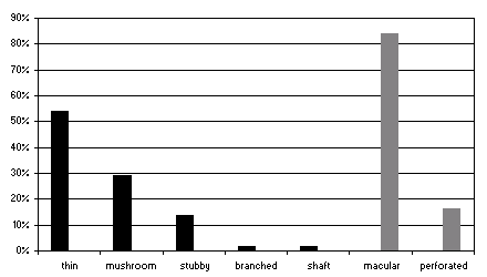
Figure 4. Distribution of Synapses.
Figure 4 shows the distribution of synapses among the various locations and shapes in stratum radiatum as measured over many adult rats. Synapses on thin spines are the most common. Most synapses have a macular shape.