Axons are functionally specialized to conduct action potentials, and structurally specialized to do so over extreme distances. Axons can attain overall lengths totaling many centimeters not just in the giraffe’s neck, but also within locally projecting, highly branched arbors in cortex. Yet, in cross section, the diameter of an axon and its synaptic contacts may be less than a micrometer. Axonal architecture thus consists of functional elements that span nearly all spatial levels of neural organization.
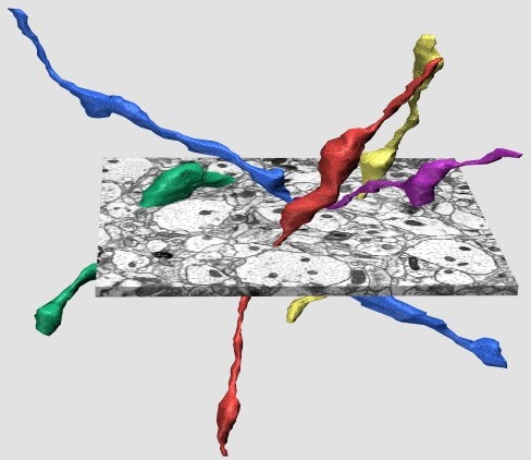
Shown here is the microscopic end of the scale: the ultrastructural appearance of individual unmyelinated axonal branches from an intracortical axonal arbor. Short portions of several axons (the tubular, colored structures) are seen passing through a slab of cerebral cortex, from stratum radiatum of area CA1 in the adult rat hippocampus. These axons were three-dimensionally reconstructed from a stack of ~100 electron micrographs.
The axon segments are ~10 micrometers in length. The synapses (not shown) occur at the swellings, also called varicosities or synaptic boutons. The tight spacing of varicosities -- one every 3 to 5 micrometers along the axon, on average -- enables a single axonal arbor to make tens of thousands of synapses. Curiously, mitochondria populate only 50% of the varicosities, raising the possibility of differences in the regulation of Ca2+ and ATP. The axonal shafts between the varicosities are extremely thin, with diameters averaging 170 nanometers -- just enough to contain a cytoskeletal scaffolding of one to several microtubules. Action potentials appear to propagate along these branches with a high degree of reliability, but at rates far slower than larger-diameter axons.
Further reading:
Shepherd GMG, Harris KM (1998) Three-dimensional structure and composition of CA3-CA1 axons in rat hippocampal slices: Implications for presynaptic connectivity and compartmentalization. The Journal of Neuroscience, 18:8300-8310. (964K PDF)