The figure below shows a section of neuropil at high magnification (red scale bar: 0.5 microns). To aid in visualization objects have been colorized. Four different axonal boutons are colorized, in blues and greens. A dendrite and its associated spine are colored pink. Another spine head, that connects to a different dendrite (not shown), is colored orange.
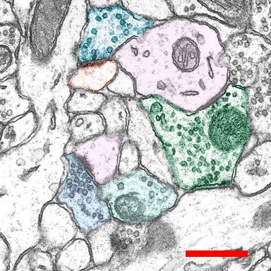
The spines each make a synapse with an axon. The synaptic regions are distinguishable by a postsynaptic density (PSD). Use the mouse to identify these and some other organelles in the image. Note that the large axon profile contains flattened vesicles and makes a different kind of synapse, a symmetric one without an enlarged PSD. These symmetric synapses are inhibitory, while those with PSDs are excitatory.