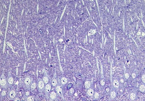
Figure 1. Semithin section from area CA1.
The stratum radiatum located next to the layer of pyramidal cells in hippocampal area CA1 is a cell body-poor, finely textured zone striated by dendritic shafts running in parallel from apices of pyramidal cells (Figure 1).
This unique felty substance, believed to be one of the most highly organized in the universe, is called neuropil. It represents a complicated spatial network comprising interconnected neuronal processes intermingled with irregularly shaped processes of astrocytic glia. Here in the hippocampus most synapses occur in neuropil. They are located mainly on dendritic spines. Synaptic neuropil is the basic constituent of the gray matter of the brain and spinal cord.
Figure 2. Electron micrograph of CA1 neuropil. Enlargement (1.2Mb GIF)
A two-dimensional electron micrograph of an ultrathin section through the neuropil (Figure 2) contains a great number of neuronal profiles represented by axons (Ax) which are forming synaptic contacts (Sy) on dendritic shafts (D) or spines (S) and are intervened by glial processes of astrocytes (Ap). Also cell bodies and processes of oligodendroglia and microglia can be sporadically present and, of course, numerous blood capillaries. To identify profiles of all these structures is a highly professional work and for analyzing their ultrastructure in ultrathin sections we recommend you consult the most detailed monography in this field, Fine Structure of the Nervous System: Neurons and Their Supporting Cells by A. Peters, S.L. Palay, and H. deF. Webster, Oxford University Press, New York and Oxford, 1991.

Figure 3. 3D reconstruction of neuropil.
When reconstructed from series of ultrathin sections into three dimensions, a spatial puzzle is obtained, representing the closest to a real view of neuropil which can be seen using up-to-date imaging methods (Figure 3).
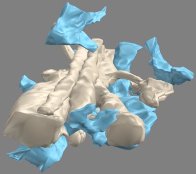
Figure 4. 3D neuropil with astrocytic processes colored pale blue.
If various structural elements from which this puzzle is built are labeled by particular colors or isolated one from another, the confusing complex architecture of the neuropil can be step by step analyzed. Astrocytic glial processes are colored in pale blue in Figure 4 leaving uncolored all processes of neural character.
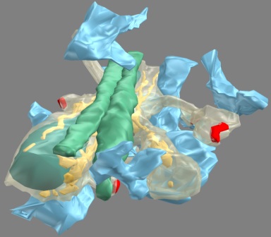
Figure 5. Color-coded 3D view of neuropil.
In Figure 5, axons were colored in green and the plasma membrane of the dendritic shaft and its spines was made semitransparent, thus enabling to visualize mitochondrion (blue-green), smooth endoplasmic reticulum (yellow) and axo-spinous synapses (red).