The following three-dimensional reconstructions are not a complete list of possible spine and synapse variations. They only illustrate the most typical examples.
Hippocampus, rat

Fig. 1: A segment of pyramidal cell dendrite from stratum radiatum (CA1) with thin, stubby, and mushroom-shaped spines. Spine synapses colored in red, shaft synapses colored in blue. The dendrite was made transparent in the lower image to enable visualization of all synapses.
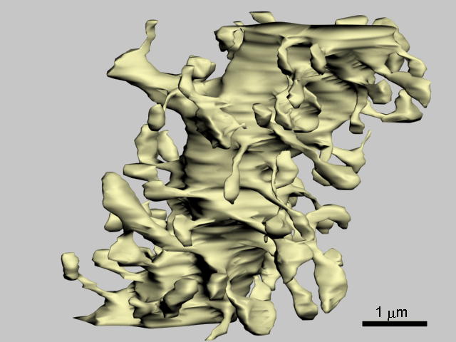
Fig. 2: Apical dendrite of pyramidal cell in stratum radiatum (CA1) with thin and mushroom-shaped spines.
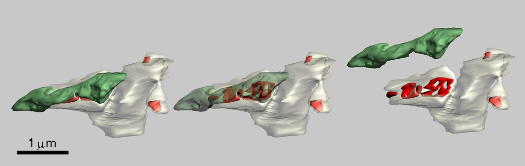
Fig. 3: A short segment of a dendrite with thin and mushroom-shaped spines, macular and perforated synapses (red) and an axon (green).
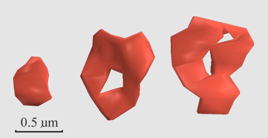
Fig. 4: Macular (A) and perforated (B, C) spine synapses. Macular synapses are located on stubby and thin spines, perforated on mushroom-shaped spines.
Neocortex, human
Dendritic spines and synapses of neocortical pyramidal cells in humans have the same appearances as those of laboratory animals.
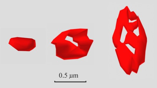
Fig. 5: Human neocortical (frontal cortex) macular and perforated spine synapses.
Cerebellar cortex, mouse
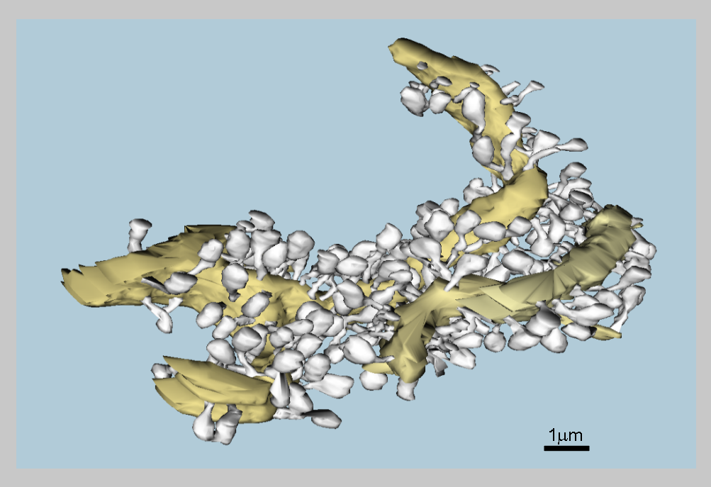
Fig. 6: Dendritic branchlets of the Purkinje cell (yellow) with numerous spines (gray).
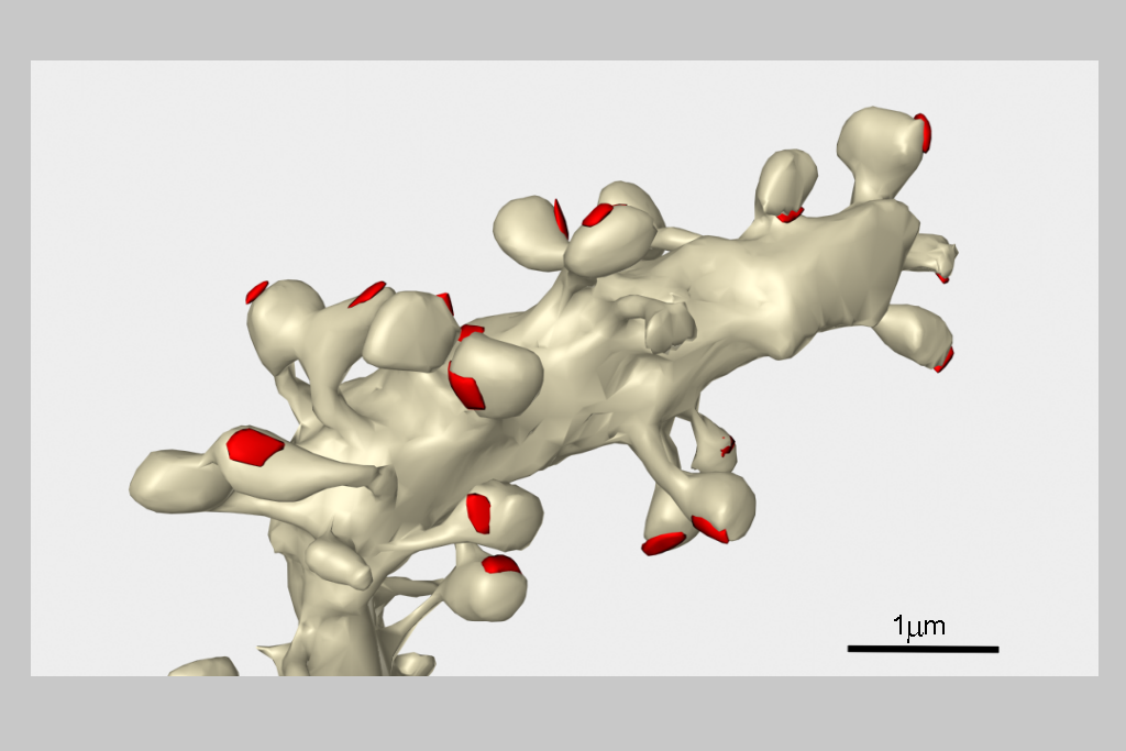
Fig. 7: Numerous club-shaped spines on a dendritic branchlet of the Purkinje cell cell in cerebellar cortex of mouse.
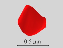
Fig. 8: Only macular synapses are located on Purkinje cell dendritic spines.
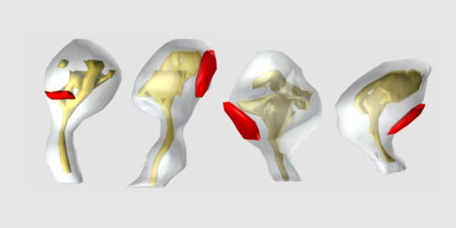
Fig. 9: Purkinje cell dendritic spines are rich in smooth endoplasmic reticulum which is, however, lacking a higher differentiation typical for the spine apparatus.
Thalamus, rat
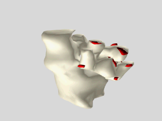
Fig. 10: Complex spines in the thalamic ventrobasal nucleus. They are associated with so called synaptic glomeruli. The synaptic glomeruli are formed in this nucleus by lemniscal giant axon terminals invaginated by ramified spines originating from proximal dendrites of thalamocortical relay neurons.
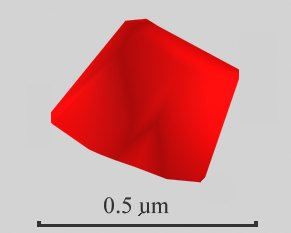
Fig. 11: Up to several macular synapses are present on one spine and several tens of synapses are present in one glomerule.
Occasionally, somatic spines and spines on axonal initial segments are also observed.
References
- Harris KM, Stevens JK (1988) Dendritic spines of rat cerebellar Purkinje cells: Serial electron microscopy with reference to their biophysical characteristics. J.Neurosci. 8:4455-4469.
- Harris KM, Jensen FE, Tsao B (1992) Three-dimensional structure of dendritic spines and synapses in rat hippocampus (CA1) at postnatal day 15 and adult ages: Implications for the maturation of synaptic physiology and long-term potentiation. J.Neurosci. 12:2685-2705.
- Peters A, Kaiserman-Abramof IR (1969) The small pyramidal neuron of the cerebral cortex. The synapses upon dendritic spines. Z. Zellforsch. Mikrosk. Anat. 100:487-506
- Spacek J, Lieberman AR (1974) Ultrastructure and three-dimensional organization of synaptic glomeruli in rat somatosensory thalamus. J. Anat. (Lond.) 117:487-515.
- Spacek J, Hartmann M (1983) Three-dimensional analysis of dendritic spines. I. Quantitative observations related to dendritic spine and synaptic morphology in cerebral and cerebellar cortices. Anat. Embryol. 167:289-310.