A neuron typically has many dendrites and one axon. The dendrites branch and terminate in the vicinity of the cell body. In contrast, axons can extend to distant targets, more than a meter away in some instances.
Dendrites are rarely more than about a millimeter long and often much shorter. Here are the dimensions of dendrites for a few types of neurons.
Neurons communicate through specialized junctions called synapses. Axons typically make synapses with other neurons through specialized enlargements near their terminals. These synapses can occur on the cell bodies or the axons of other neurons, but most frequently they occur on dendrites. Thus, the dendrites of a neuron provide a surface for receiving synaptic inputs from other neurons.
The arbor formed by the dendrites of a neuron often has a characteristic shape as determined by the spatial domains into which the dendrites ramify. Here are some examples of patterns of dendritic arborization.
Dendrites of some neurons are smooth, tapered processes, such as in motor neurons of the spinal cord. Other neurons exhibit enlargements, protrusions, or other structural specializations along dendrites, or frequently, at the ends of dendrites. These structures are often sites of synaptic contact and therefore can be referred to as synaptic specializations. Here are some examples of synaptic specializations of dendrites.
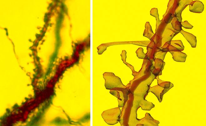
Spiny dendrites from hippocampal pyramidal neuron.
Left: Light microscope image. Right: Reconstruction from serial electron microscopy.
Perhaps the most common synaptic specialization of dendrites is that which Spanish anatomist Ramon y Cajal referred to as "espinas", since they resembled the thorns on a flower stem. These spines are frequent on the dendrites of the principal cells of most brain regions, notably on the pyramidal cells of cerebral cortex and the Purkinje cells of the cerebellar cortex. For these cells, more than 90% of their excitatory synapses occur on dendritic spines. Therefore, spines may play an important role in learning and memory.
Simple spines are very small, often less than 1 micron in diameter. This makes them difficult to study through light microscopy. Electron microscopy must be used to determine the geometry of spines. Here are some dimensions of simple dendritic spines as determined through serial electron microscopy.
Adapted from:
Fiala J.C., Harris K. M. (1999) Dendrite Structure. In Dendrites (eds. G. Stuart, N. Spruston, M. Häusser), Oxford University Press.