Fundamental to understanding the mechanisms of synaptic transmission in the central nervous system is knowing the dimensions and connectivity of synapses and the changes in structure that occur during development and plasticity. Improved computer-based quantitative analysis of brain anatomy at the ultrastructural level, especially the analysis of synaptic structure and connectivity may clarify many of the issues in a way that no other technique can.
Most excitatory synapses in the adult brain are located on the bulbous heads of dendritic spines which occur in a variety of shapes and sizes. During development, before the expression of spines, cortical dendrites exhibit very long, thin processes, often without a bulbous head, called filopodia. As the cortex matures, these dendritic filopodia disappear and spines emerge.
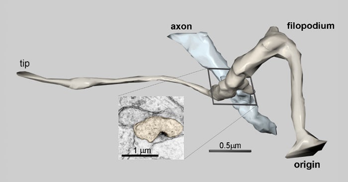
Serial section electron microscopy and three-dimensional analysis were used to determine the role of dendritic filopodia in the genesis of excitatory synaptic contacts and dendritic spines in hippocampal area CA1 in developing rats. The analysis revealed that numerous dendritic filopodia formed synaptic contacts with axons and with filopodia extending from axons. Recent studies have suggested that these filopodia are involved in locating presynaptic targets and consolidating them into stable synapses. This figure shows a three-dimensional reconstruction of a dendritic filopodium created from the serial electron micrographs. The inset uses an electron micrograph to illustrate the corresponding synaptic connection between the filopodia and the adjacent axon.
Reference:
Fiala, J.C., Feinberg, M., Popov, V. and Harris, K. M. (1998) Synaptogenesis via dendritic filopodia in developing hippocampal area CA1. J. Neuroscience, 18(21):8900-8911. (PDF)