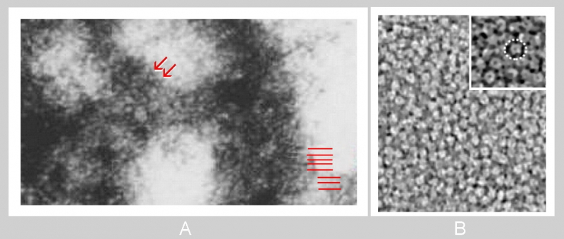
Fig. 1.6.45. A detail from Fig. 1.6.36. A tangentional section through axo-dendritic synapse showing structures supposed to represent extracellular domains of glutamate receptors (A - arrows) and scaffolding filaments of a postsynaptic density (red lines). Compare to acetylcholine receptors revealed by cryo-electron microscopy (courtesy of PNAS and Zuber et Unwin, 2013; doi 10.1073/pnas.1301277110) (B). Diameters of putative (A) and accredited (B) receptors are around 10 nm. (Neocortex, mouse (A), and neuromuscular junction (B), Torpedo marmorata.)