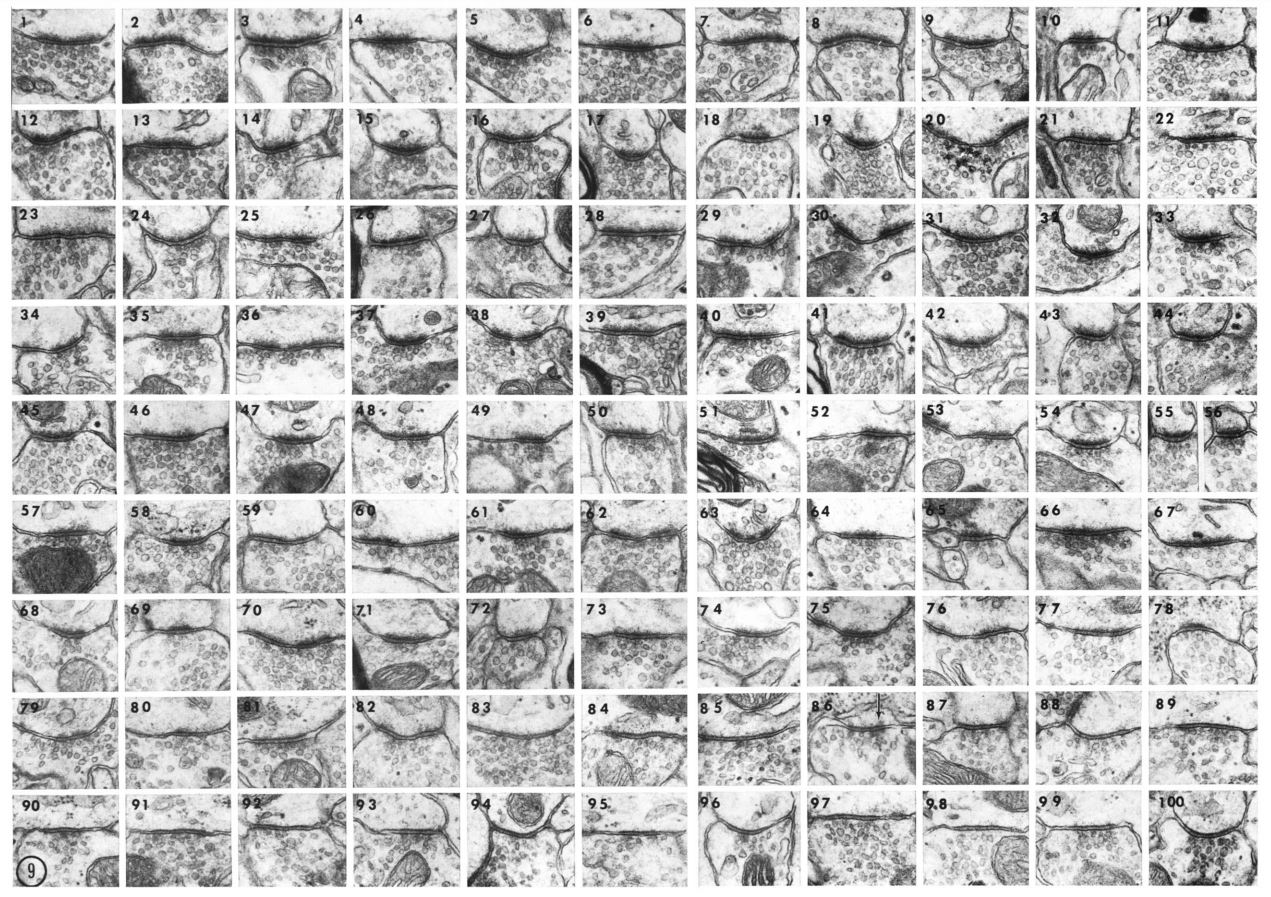
A montage of 100 synapses illustrating differences in opacity of the postsynaptic density (PSD) and shape of synaptic vesicles. The synapses are displayed in approximate order of decreasing opacity. For synapses 1-56, it is impossible to discern an exact order since the PSDs appear similar. For image 57 and higher, the density decreases in thickness and the vesicles appear more elongate by image 83. For purposes of classification, Colonnier considered images 1-79 to represent asymmetric synapses whereas synapses 83-100 were identified as symmetric. He concluded that these two classifications were sufficient since a proportion of synapses failed to appear morphologically "transitional" between the asymmetric and symmetric categories (x 17000).
Reproduced from: Colonnier M. (1968) Synaptic patterns on different cell types in the different laminae of the cat visual cortex. An electron microscope study. Brain Res., Jul 9(2):268-87.