
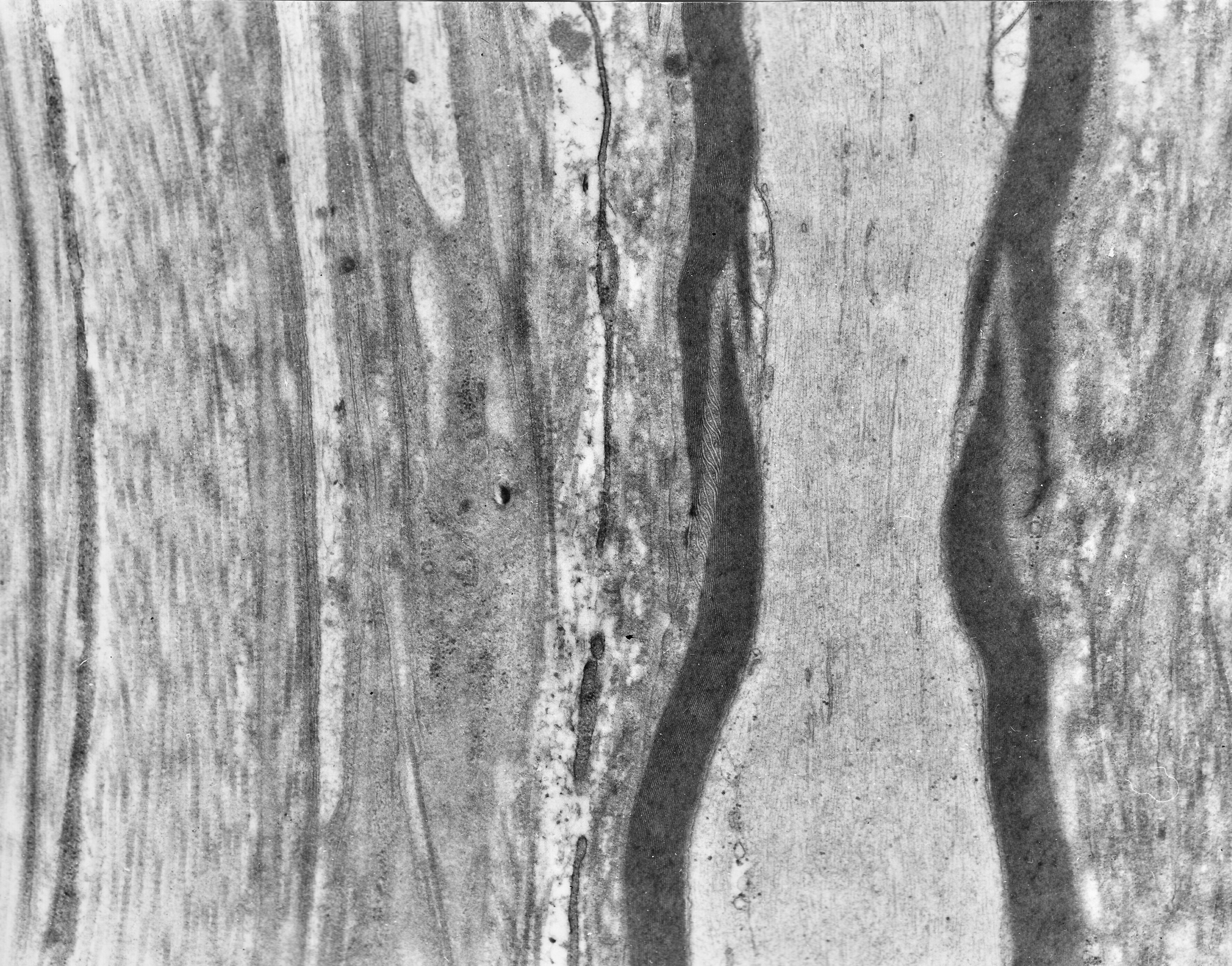
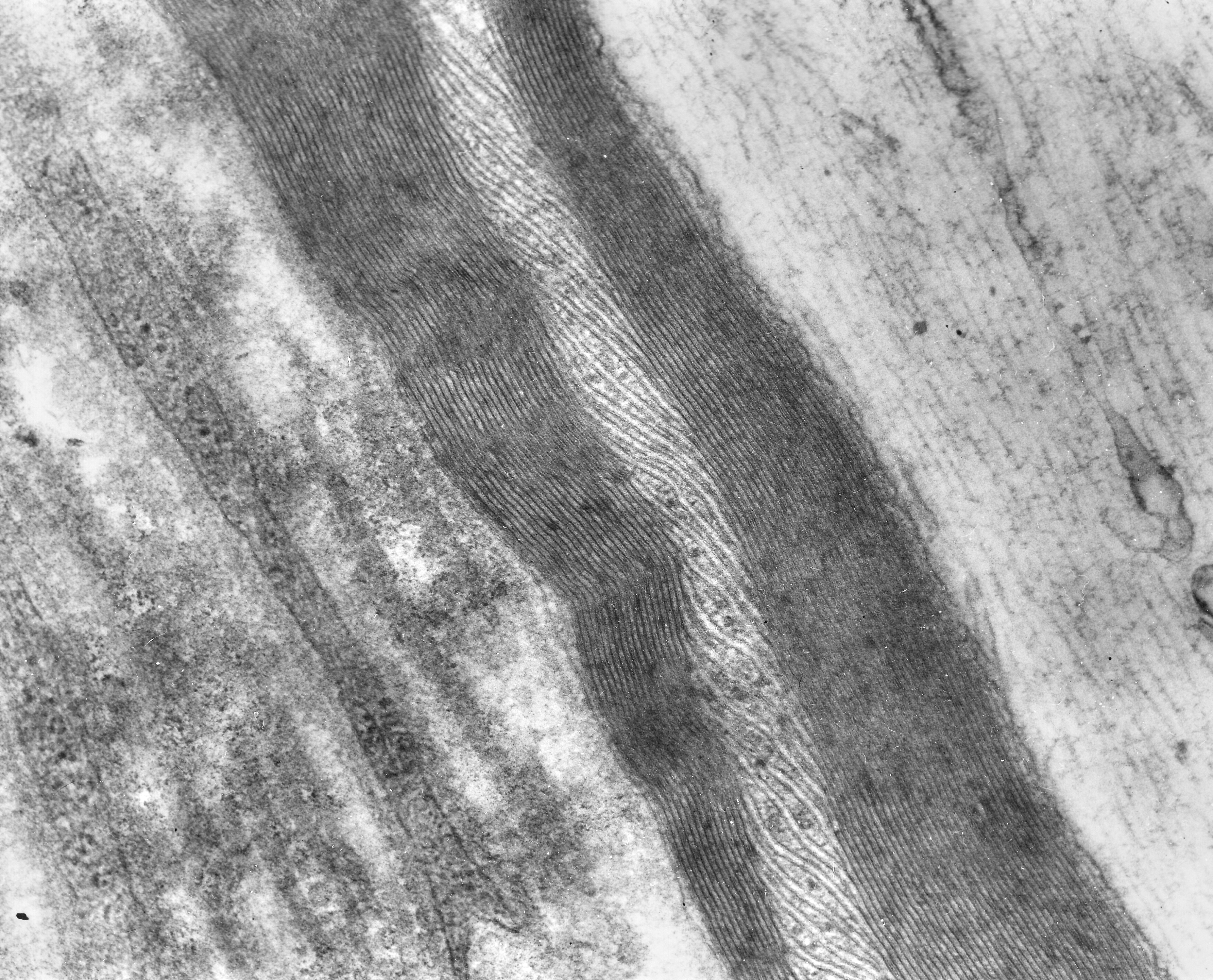
Fig. 5.15. Upper figure: Light microscopy of the funnel-shaped Schmidt-Lanterman incisures as they appear in Luxol Fast Blue-stained longitudinal paraffin section of peripheral nerve. (Human, peripheral nerve.) Lower figure: Longitudinally cut myelinated axons with Schmidt-Lanterman incisures (arrows). Scale = 1 µm. (Human, sural nerve.) Download the high resolution image (b); Download the high resolution image (c).