
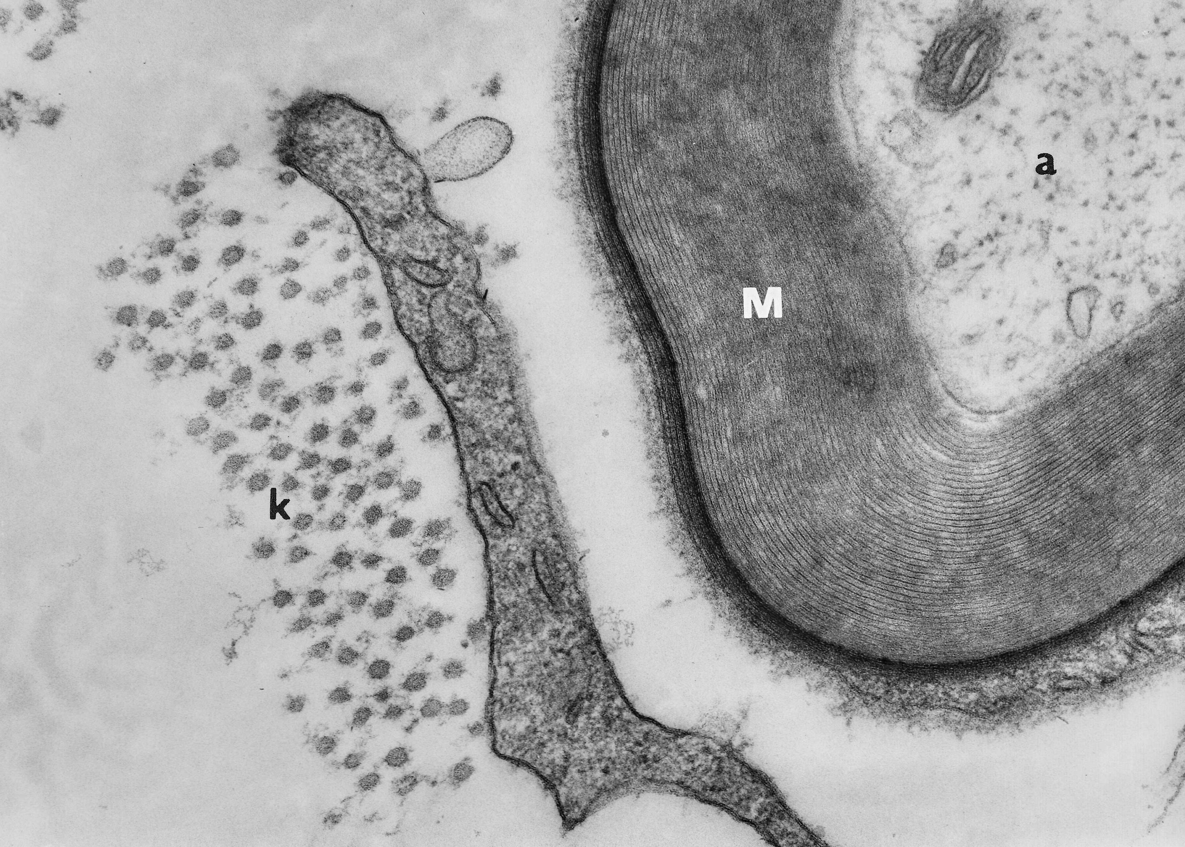
Fig. 5.06. Myelinated axon (M, a) and thin fibrocyte process (f) with a bunch of collagen fibres (c) in the endoneurial space. Basal lamina is marked by arrow. Scale = 200 nm. (Rat, trigeminal nerve.) Download the high resolution image.
Fig. 5.06. Myelinated axon (M, a) and thin fibrocyte process (f) with a bunch of collagen fibres (c) in the endoneurial space. Basal lamina is marked by arrow. Scale = 200 nm. (Rat, trigeminal nerve.) Download the high resolution image.