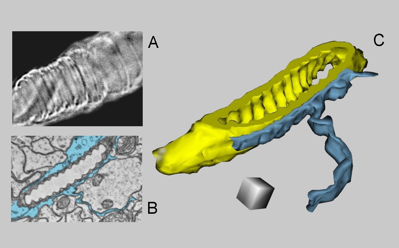
Fig. 3.23. A: Tracheole of an insect (light microscopy). B: An ultrastructural appearance of a tracheole in the antennal lobe of Drosophila brain in one section of the series, from which the 3D reconstruction was made (C). Glial processes (blue) surround the tracheole. Scale cube = 0.2 µm per side. (Antennal lobe, Drosophila brain.) Series from Zheng et al. 2018 used for the reconstruction.