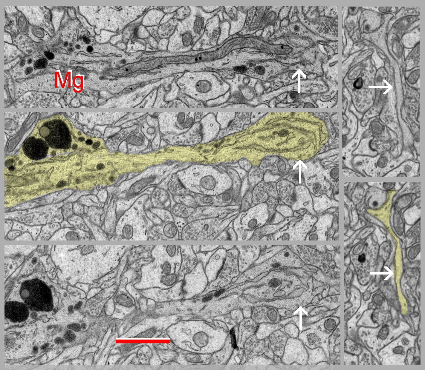
Fig. 2.4.13. Three consecutive serial sections of a microglial cell (Mg). A thick process (arrows) protrudes from the cell body containing numerous electron-dense lysosomes. Thin peripheral microglial processes are visible in the right images (arrows) and can only be distinguished in serial sections from very similar dendritic filopodia (cf. Figs. 1.4.3.05, 1.4.3.07, 1.4.3.08). Download the high resolution image. Scale bar = 1 µm. (Hippocampus, rat.)