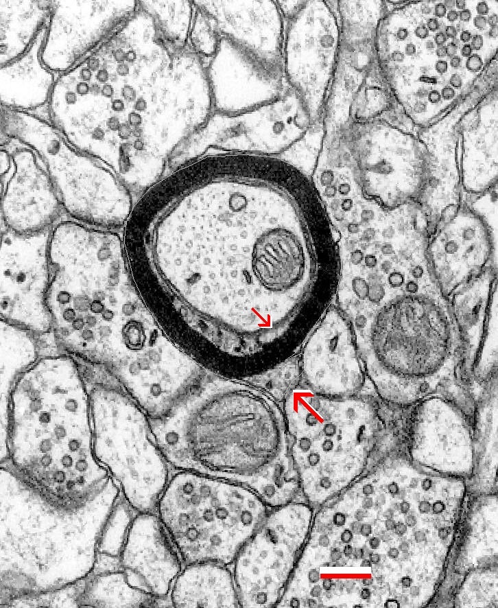
Fig. 2.3.16. Myelinated axon in an ideally oriented transverse section. The outer (larger arrow) and inner (smaller arrow) tongues of the oligodendroglial process are clearly visible. Scale bar = 0.2 µm. (Hippocampus, rat.)
Fig. 2.3.16. Myelinated axon in an ideally oriented transverse section. The outer (larger arrow) and inner (smaller arrow) tongues of the oligodendroglial process are clearly visible. Scale bar = 0.2 µm. (Hippocampus, rat.)