
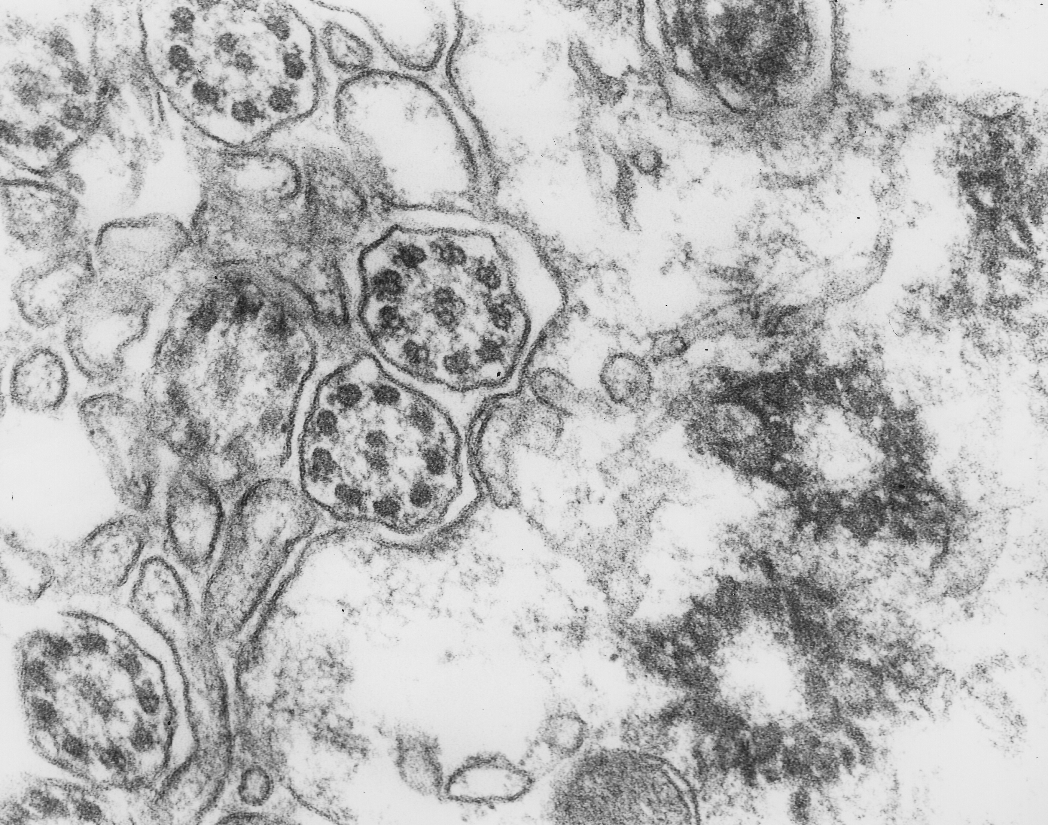
Fig. 2.2.06. Transversally cut ependymal cilia (red arrow) and ciliary basal bodies in the subsurface region of the ependymal cell (blue arrow). Scale = 100 nm. (Mouse, 4th ventricle.) Download the high resolution image.
Fig. 2.2.06. Transversally cut ependymal cilia (red arrow) and ciliary basal bodies in the subsurface region of the ependymal cell (blue arrow). Scale = 100 nm. (Mouse, 4th ventricle.) Download the high resolution image.