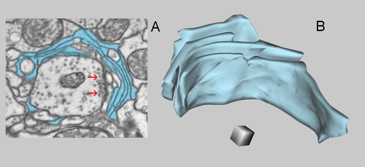
Fig. 2.1.44. A: Neurite from the antennal lobe of a Drosophila brain (intepreted as axon) is surrounded by glial lamellae (blue). Note axo-axonal synapses (arrows), the postsynaptic densities in profiles proved to be thin projections from axonal boutons, are inapparent. B: A partial 3D reconstruction of the same glial lamellae. Scale cube = 0.2 µm per side. Cf. Fig.1.6.23. (Antennal lobe, Drosophila brain.) Series from Zheng et al. 2018 used for the reconstruction.