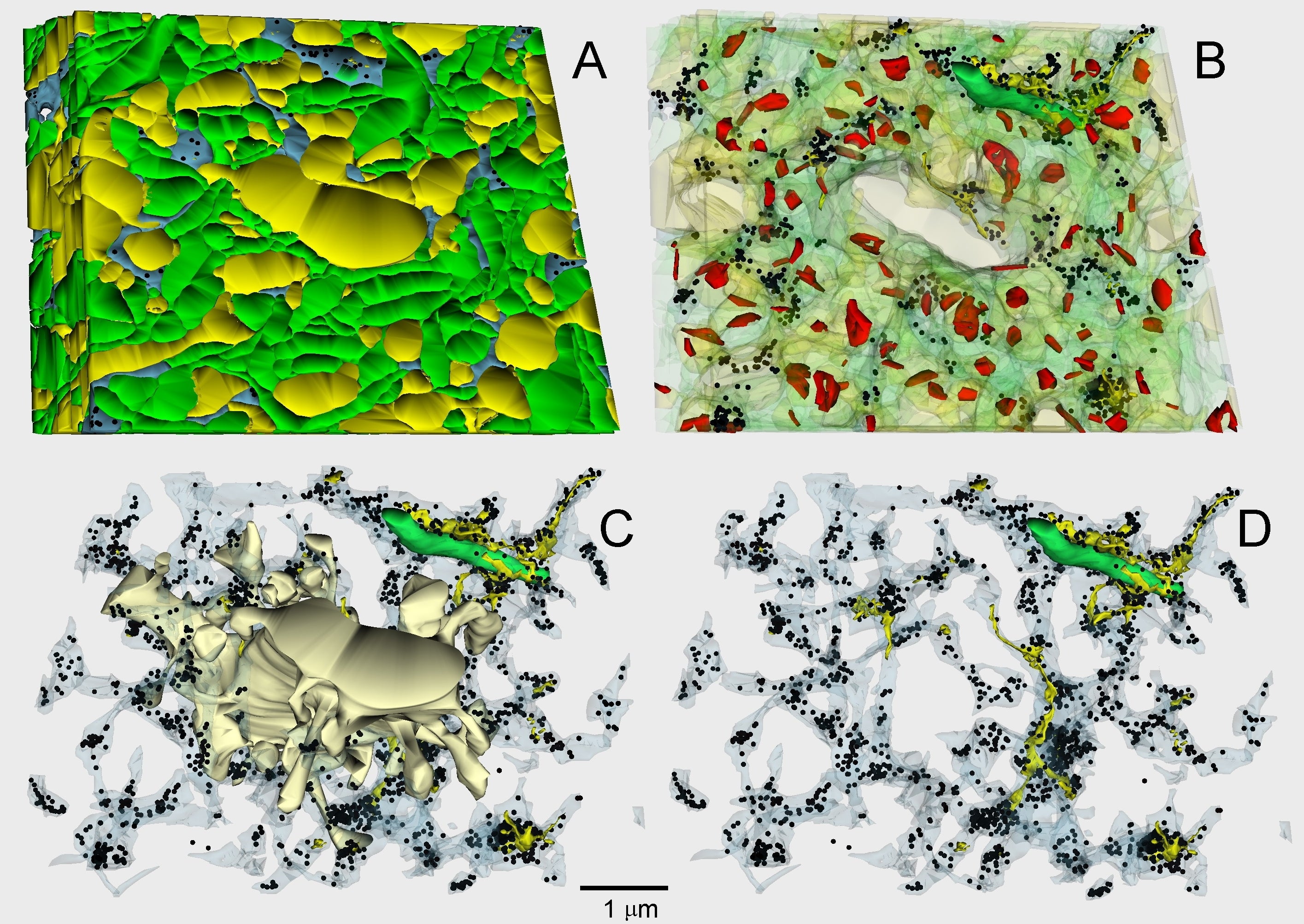
Fig. 2.1.43. Three-dimensional reconstruction from 20 (A, B) or 40 (C, D) sections of a volume of the hippocampal neuropil shown in Figs. 1.5.32 and 1.9.27. Plasma membranes of axons (green), dendrites (yellow), and astrocytic glia (blue) in A, made transparent in B to see synapses (red). In C, all neurites were omitted apart from an apical dendrite. In transparent astrocyte processes, mitochondrion (green), endoplasmic reticulum (yellow), and glycogen particles (black) are apparent (C). Mitochondria was present in the widest process only; practically no endoplasmic reticulum or greater groups of glycogen particles were present in peripheral lamellar astrocyte processes (D). No synapse-specific distribution of glycogen was observed in our sample. Scale = 1 µm. (Rat, hippocampus.)