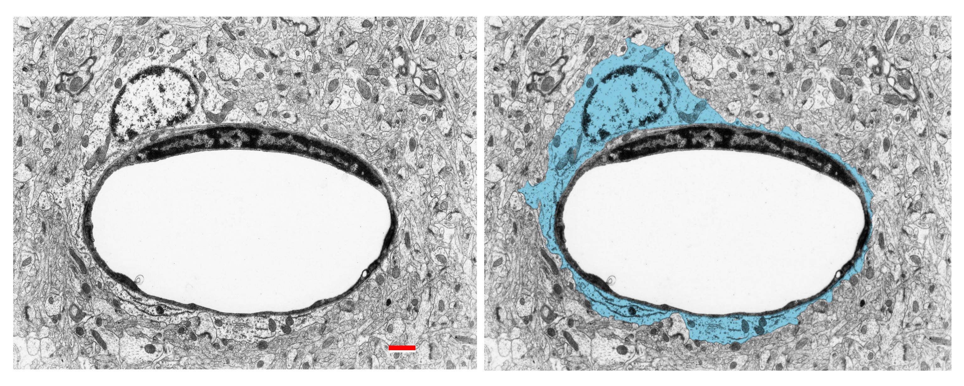
Fig. 2.1.38. Pericapillary located astrocyte (marked in blue). Its thin flattened processes completely ensheathe the capillary wall. Download the high resolution image. Scale = 1 µm. (Human, epitumorous neocortex.)
Fig. 2.1.38. Pericapillary located astrocyte (marked in blue). Its thin flattened processes completely ensheathe the capillary wall. Download the high resolution image. Scale = 1 µm. (Human, epitumorous neocortex.)