
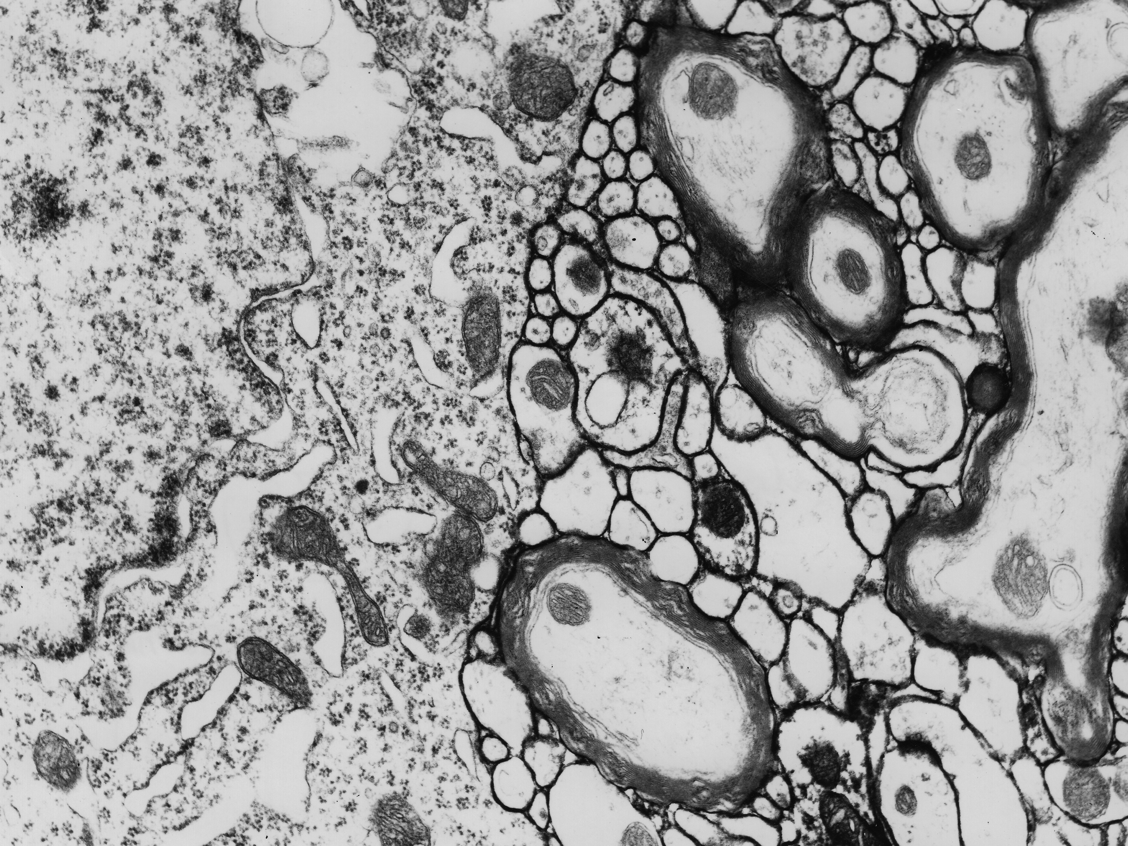
Fig. 1.8.02. Narrow intercellular clefts between elements of the neuropil are marked with an electron-dense ruthenium red in this electron micrograph (arrow). P - neuronal perikaryon. Scale = 300 nm. (Rat, thalamus.) Download the high resolution image.