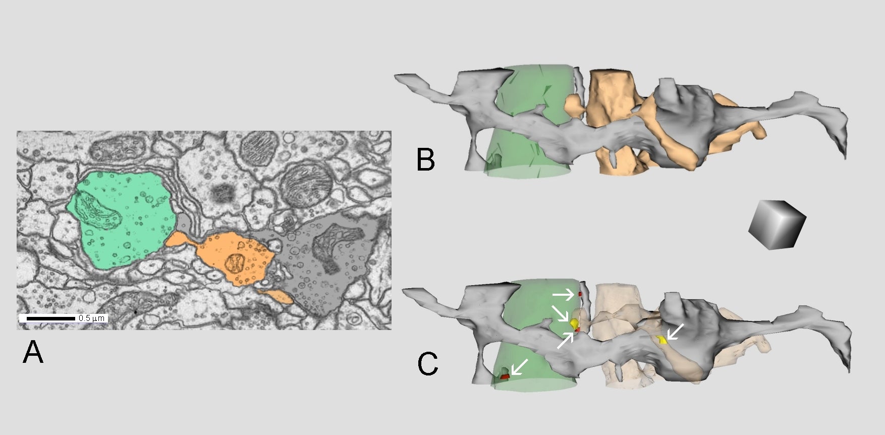
Fig. 1.5.39. A: Neuropil from antennal lobe of a Drosophila brain. B: 3D reconstruction of three neurites interpreted as axons from the same region. C: Axo-axonal synapses are marked by arrows: the brown axon makes synapse with the grey one (yellow), and both the brown and grey axons make synapses with the green one (yellow, red). Scale cube = 0.5 µm per side. (Antennal lobe, Drosophila brain.) Series from Zheng et al. 2018 used for the reconstruction.