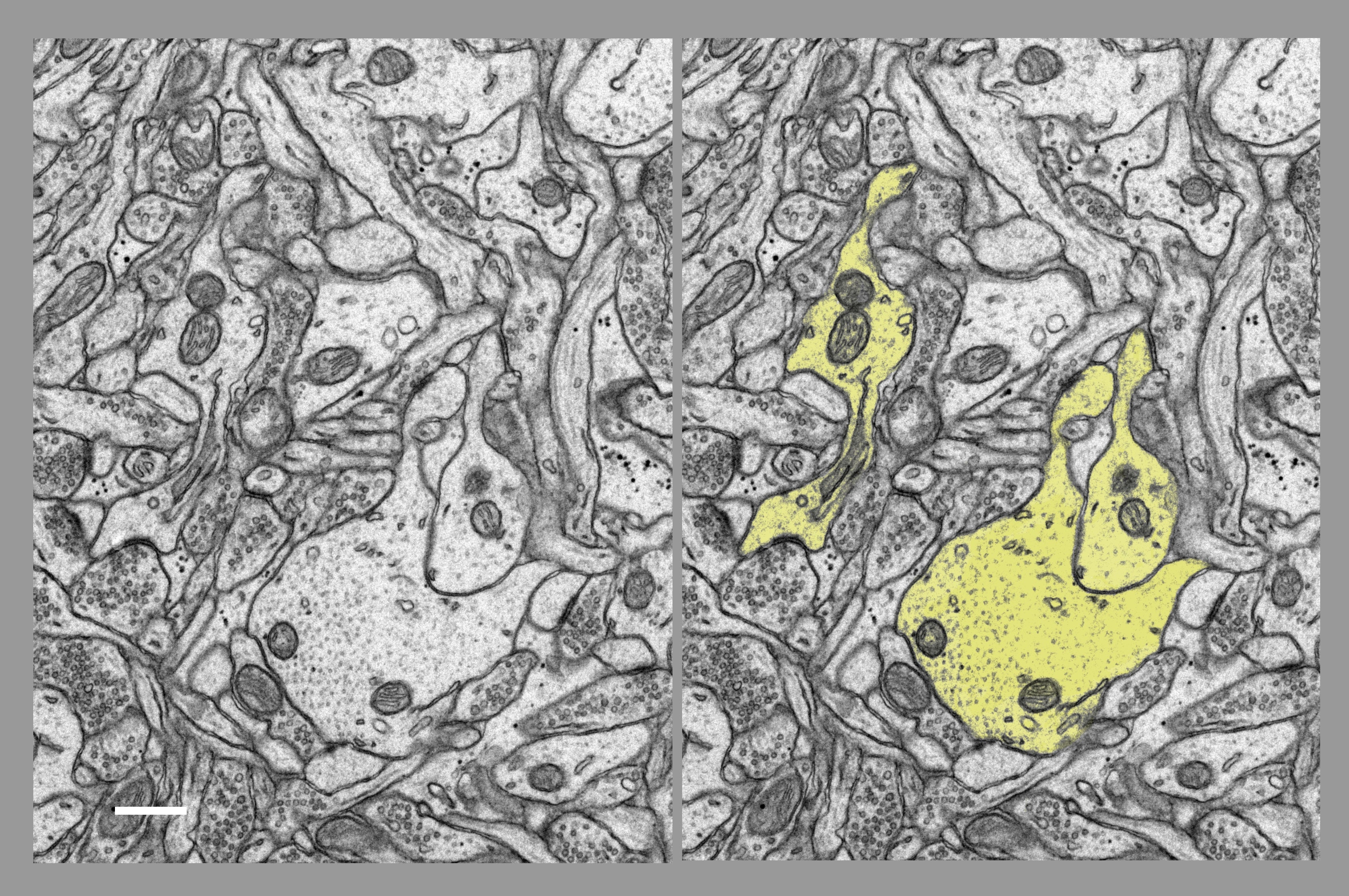
Fig. 1.4.1.62. Group of transversally sectioned dendrites (yellow) coincidentally (and ideally) showing dendritic spines with their necks, heads and synaptic contacts. Download the high resolution image. Scale bar = 0.5 µm. (Hippocampus, rat.)
Fig. 1.4.1.62. Group of transversally sectioned dendrites (yellow) coincidentally (and ideally) showing dendritic spines with their necks, heads and synaptic contacts. Download the high resolution image. Scale bar = 0.5 µm. (Hippocampus, rat.)