
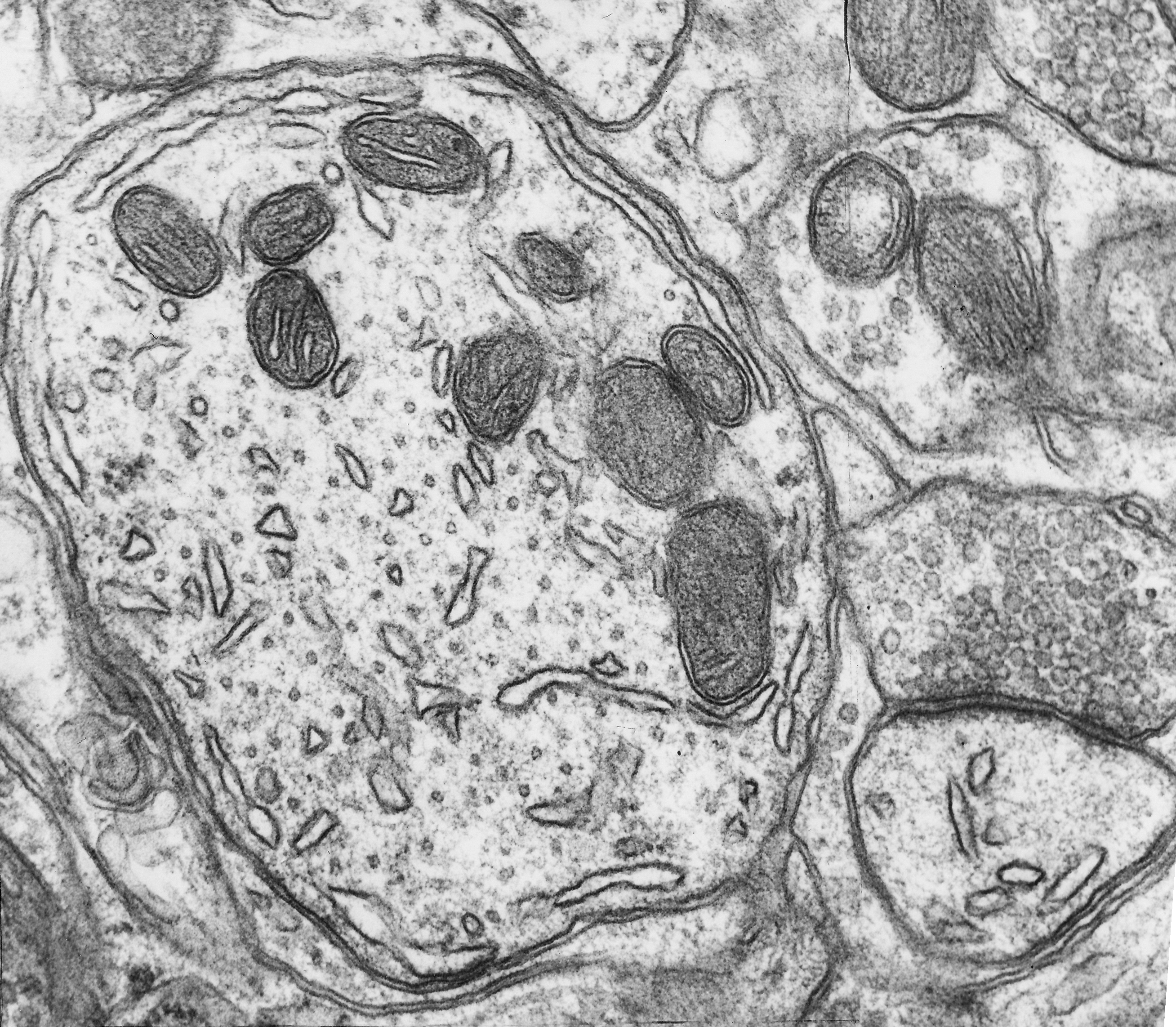
Fig. 1.4.04. Secondary dendrite of a Purkinje cell. Microtubules (red arrow) and smooth endoplasmic reticulum (blue arrow) are shown transversally sectioned. Scale = 200 nm. (Mouse, cerebellar cortex.) Download the high resolution image.
Fig. 1.4.04. Secondary dendrite of a Purkinje cell. Microtubules (red arrow) and smooth endoplasmic reticulum (blue arrow) are shown transversally sectioned. Scale = 200 nm. (Mouse, cerebellar cortex.) Download the high resolution image.