
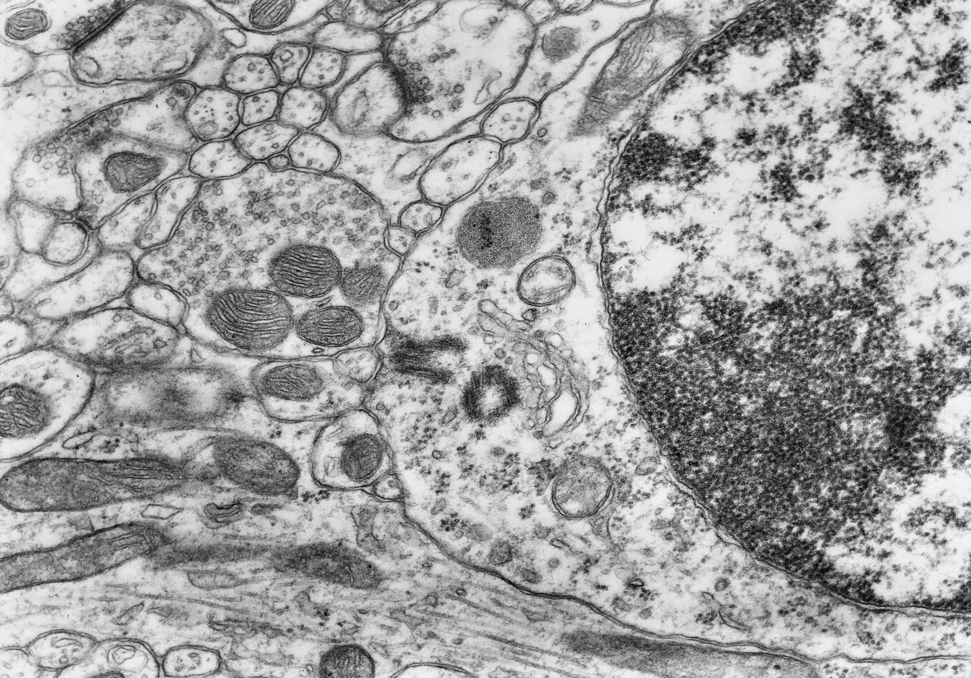
Fig. 1.1.7.01. Two centrioles perpendicular to each other (red arrow) in the cerebellar granule cell. N - nucleus, blue arrow - secondary lysosome. Scale = 300 nm. (Mouse, cerebellar cortex). Download the high resolution image.
Fig. 1.1.7.01. Two centrioles perpendicular to each other (red arrow) in the cerebellar granule cell. N - nucleus, blue arrow - secondary lysosome. Scale = 300 nm. (Mouse, cerebellar cortex). Download the high resolution image.