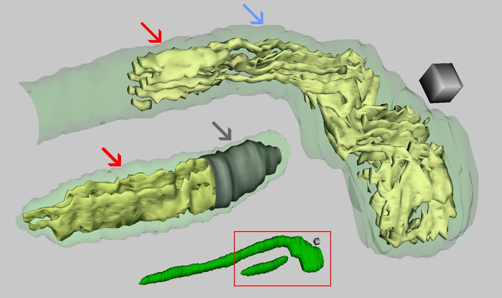
Fig. 1.1.4.13. 3D reconstruction of two axonal mitochondria. Flat cristae (red arrows) turn into tubular channels in places (blue arrow). Inner mitochondrial membrane (grey arrow) was omitted. Scale cube = 0.1 μm per side. (Mouse, cerebellum.)
Fig. 1.1.4.13. 3D reconstruction of two axonal mitochondria. Flat cristae (red arrows) turn into tubular channels in places (blue arrow). Inner mitochondrial membrane (grey arrow) was omitted. Scale cube = 0.1 μm per side. (Mouse, cerebellum.)