A chemical synapse is a modified intermediate intercellular junction specialized for a chemical transmission of electrical signals. However, a great number of spinous synapses (at least 77% of perforated synapses) are inhomogeneous and contain undifferentiated vesicle-free portion having character of punctum adherens. As proved by recent immunocytochemical studies, adhesion molecules (cadherins – Fig. 3, catenins, neural cell adhesion molecule - NCAM) are concentrated in these areas. These molecules are significant not only in a processes of differentiation of neural tissue and synaptogenesis, but they are also involved in the synaptic plasticity, inclusive of LTP.
We found an intimate relationship between the spine apparatus and the synapse-associating punctum adherens-like nascent zone: a granulofilamentous material radiates from the inner dense plates of the spine apparatus into the punctum. Our suggestion is that the spine apparatus can synthesize proteins designed for extending synapse (Fig. 1).
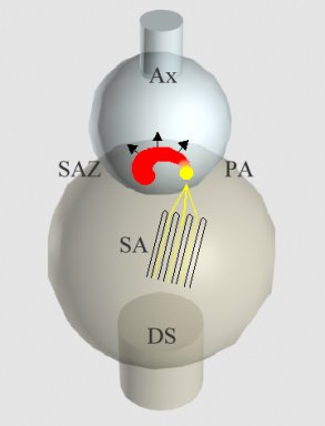
Fig. 1: A model of our suggestion how a synapse can enlarge in the process of synaptic plasticity. Ax - axon terminal; DS - dendritic spine; SA - spine apparatus; a material of the inner dense plate - in yellow; PA - punctum adherens-like nascent zone, free of synaptic vesicles; SAZ - the extending synaptic active zone.
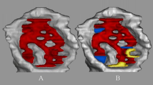
Fig. 2: The hippocampal perforated spine synapse as it looks under the classical view of homogenious ultrastructure (A) and inhomogeneous ultrastructure (B) containing vesicle-free adherent puncta (yellow), vesicle-free transitional zones (nascent zones - blue) and vesicle-associated synaptic active zone (red). See Spacek and Harris, 1998 and Bell et al., 2014, for details.
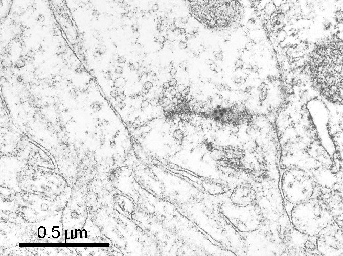
Fig. 3: Catenin marked with immunogold particles in the punctum adherens-like nascent zone. (Human, cerebral cortex.)
Apart from the punctum adherens accompanying the synapse, numerous dendritic spines, especially those of mushroom-shaped type with perforated synapse, possses another punctum adherens fixing an astrocytic process to the spine (Fig. 3). The astrocytes play an important role in maintainance of ion and perhaps also mediator homeostasis in surroundings of synapse.
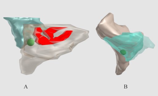
Fig. 4: A punctum adherens (green) fixing an astrocyte process (pale blue) to a mushroom-shaped dendritic spine possesing a perforated synapse (red). The spine (A) and astrocyte (B) made semitransparent to enable the punctum to be visible.
Extremely elaborated net-like vesicle-free adhesion junctions associated with synapses are formed between dendrites of thalamocortical projection neurons and giant axon terminals in synaptic glomerules of thalamic nuclei (Fig. 5).
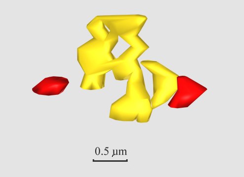
Fig. 5: Reconstruction of extensively elaborated net-like vesicle-free adhesion junctions (yellow) associated with synaptic active zones (red) between a dendrite of thalamocortical relay neuron and a lemniscal giant axon terminal in thalamic ventrobasal nucleus of rat.
References
- Bell ME, Bourne JN, Chirillo MA, Mendenhall JM, Kuwajima M, Harris KM (2014) Dynamics of nascent and active zone ultrastructure as synapses enlarge during long-term potentiation in mature hippocampus. J. Comp. Neurol. 522:3861-3864.
- Colman DR (1997) Minireview and hypothesis. Neurites, synapses, and cadherins reconciled. Molecular and Cellular Neuroscience 10:1-6.
- Fannon AM, Colman DR (1996) A model for central synaptic junctional complex formation based on the differential adhesive specificities of the cadherins. Neuron 17:423-434.
- Lieberman AR, Spacek J (1997) Filamentous contacts: the ultrastructure and three-dimensional organization of specialized non-synaptic interneuronal appositions in thalamic relay nuclei. Cell Tiss. Res. 288:43-57.
- Martin KC, Kandel ER (1996) Cell adhesion molecules, CREB, and the formation of new synaptic connections. Neuron 17:597-570.
- Spacek J (1985b) Relationships between synaptic junctions, puncta adhaerentia and the spine apparatus at neocortical axo-spinous synapses. Anat. Embryol. 173:129-135.
- Spacek J, Harris KM (1997) Three-dimensional organization of smooth endoplasmic reticulum in hippocampal CA1 dendrites and dendritic spines of the immature and mature rat. J.Neurosci. 17:190-203.
- Spacek J, Harris KM (1998) Three-dimensional organization of cell adhesion junctions at synapses and dendritic spines in area CA1 of the rat hippocampus. J.Comp.Neurol. 393:58-68.
- Uchida N, Honjo Y, Johnson KR, Wheelock MJ,Takeichi M (1996) The catenin/cadherin adhesion system is localized in synaptic junctions bordering transmitter release zones. J.Cell Biol. 135:767-779.
- Ventura R, Harris KM (1999) Three-dimensional relationships between hippocampal synapses and astrocytes. J Neuroscience 19(16):6897-6906.