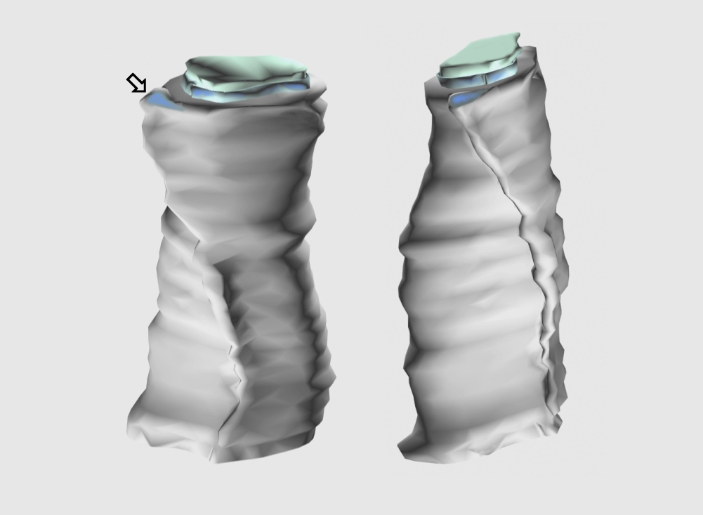
Fig. 2.3.1.10. Three-dimensional reconstruction of myelinated axon seen from different viewpoints. The axon itself is colored in pale green, oligodendroglial inner and outer cytoplasmic tongues are pale blue. Arrow is directed to the outer tongue which forms the first turn of myelin sheath. (Rat, hippocampus.) Download the high resolution image.