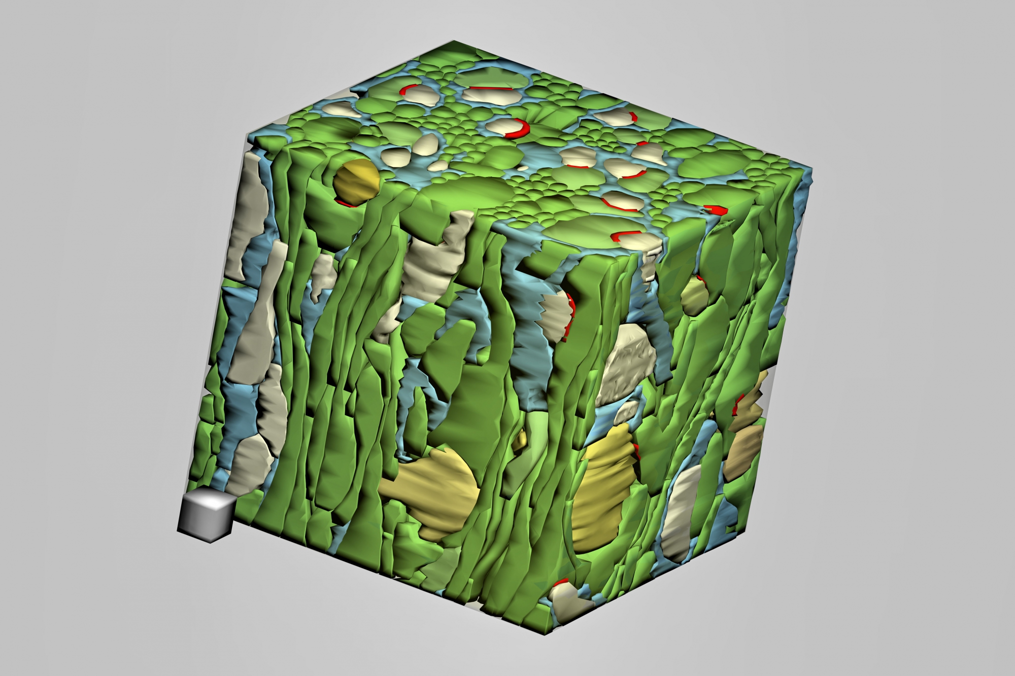
Fig. 1.9.22. Densely reconstructed 2 x 2 x 2 µm volume of neuropil from the molecular zone of the cerebellar cortex. Axons – green, dendritic branches and spines – yellow and ochre, synapses – red, astroglia – blue. (FIB-SEM series used for the 3D reconstruction provided by Lich, Wall and Knott, 2008.) Scale cube = 0.2 µm per side. (Mouse, cerebellar cortex.)