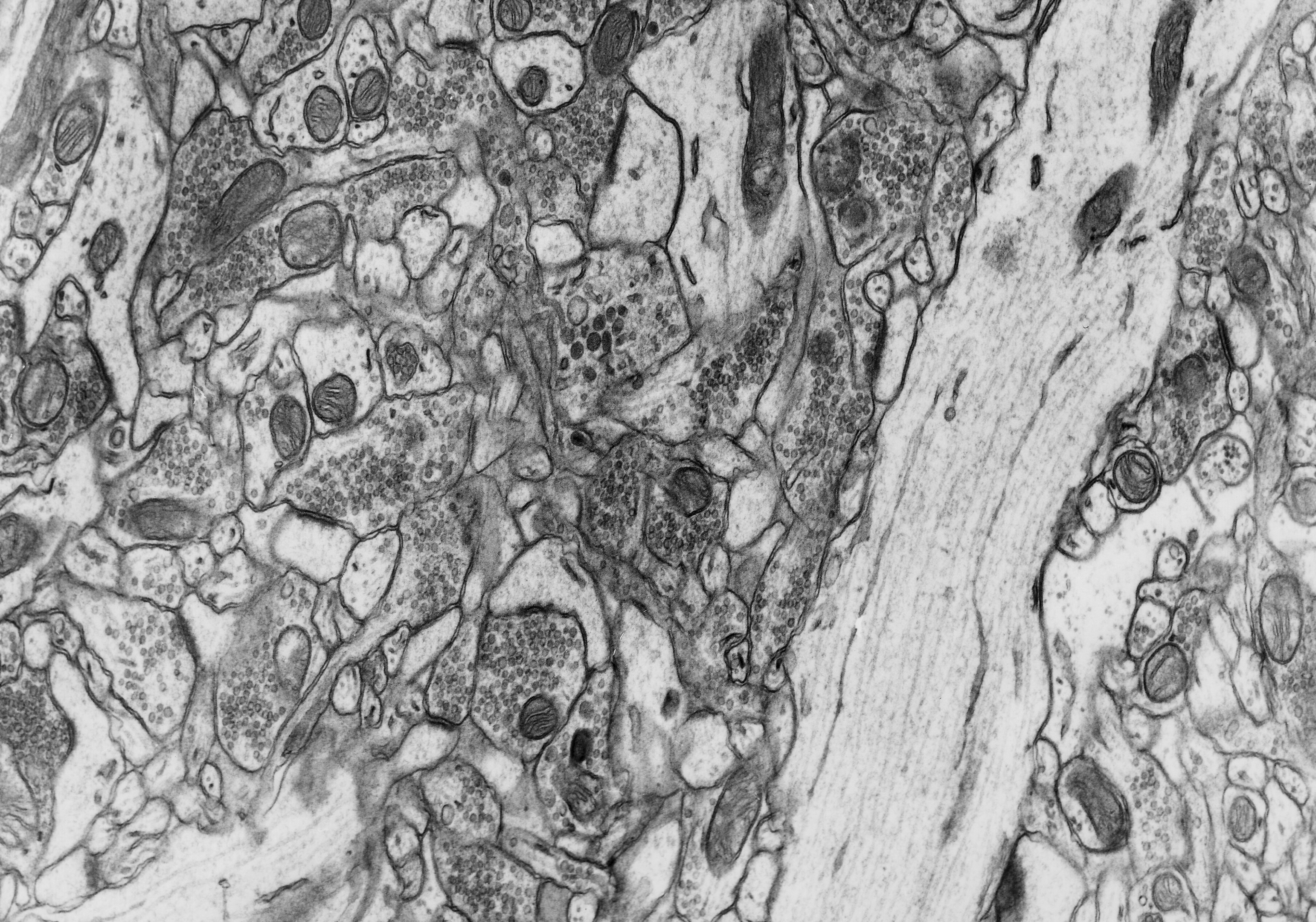
Fig. 1.8.01. The typical cortical neuropil to the left of a dendrite (D) of a pyramidal cell. Scale = 1 µm. (Rat, hippocampus.) Download the high resolution image.
Fig. 1.8.01. The typical cortical neuropil to the left of a dendrite (D) of a pyramidal cell. Scale = 1 µm. (Rat, hippocampus.) Download the high resolution image.