
Fig. 1.4.1.01. Stubby spine
Fig. 1.4.1.02. Thin spine
Fig. 1.4.1.03. Mushroom spine with spine apparatus
Fig. 1.4.1.04. Mushroom spine
Fig. 1.4.1.05. Mushroom spines
Fig. 1.4.1.06. Spine with endoplasmic reticulum and punctum adherens
Fig. 1.4.1.07. Mushroom spine with spinule
Fig. 1.4.1.08. Purkinje cell spines
Fig. 1.4.1.09. Mushroom spine in 3D
Fig. 1.4.1.10. Purkinje cell branchlet with spines
Fig. 1.4.1.11. Pyramidal cell somatic spine
Fig. 1.4.1.12. Somatic spine in 3D
Fig. 1.4.1.13. Freeze fracture of spine
Fig. 1.4.1.14. Dendritic spines from the brain of a fish
Fig. 1.4.1.15. Synaptic glomerule in thalamic ventrobasal nucleus in 3D
Fig. 1.4.1.16. Spiny dendrite in 3D
Fig. 1.4.1.17. Detail of spiny dendrite in 3D
Fig. 1.4.1.18. Endoplasmic reticulum and spine apparatus in 3D
Fig. 1.4.1.19. Spine apparatus in 3D
Fig. 1.4.1.20. Purkinje cell dendritic spines
Fig. 1.4.1.21. Spine apparatus in mushroom spine
Fig. 1.4.1.22. Synapse shapes in cerebral cortex
Fig. 1.4.1.23. Branched dendritic spine
Fig. 1.4.1.24. Polyribosomes in head and neck of spine
Fig. 1.4.1.25. Actin microfilaments in spine neck
Fig. 1.4.1.26. Polyribosome and spine apparatus in spine neck
Fig. 1.4.1.27. Polyribosome and spine apparatus in spine head
Fig. 1.4.1.28. Serial sections of complex spine filled with polyribosomes
Fig. 1.4.1.29. Complex spine filled with polyribosomes in 3D
Fig. 1.4.1.30. Spine and shaft synapses
Fig. 1.4.1.31. 3D of complex dendritic spines in thalamus
Fig. 1.4.1.32. Dendritic spine making synapse with two axon terminals
Fig. 1.4.1.33. 3-D reconstruction of dendritic spines of cerebellar Purkinje cell with flip
Fig. 1.4.1.34. 3-D reconstruction of dendritic spines of cerebellar Purkinje cell
Fig. 1.4.1.35. 3-D reconstruction of spine apparatus in spine neck
Fig. 1.4.1.36. Serial sections of spine apparatus
Fig. 1.4.1.37. 3D reconstruction of spine apparatus
Fig. 1.4.1.38. 3D reconstruction of astrocytic processes covering dendritic spines
Fig. 1.4.1.39. 3D reconstruction of dendritic spinules invading astrocyte processes
Fig. 1.4.1.40. Free postsynaptic density in dendritic spine
Fig. 1.4.1.41. A dendrite in 3D reconstruction and in Golgi impregnation
Fig. 1.4.1.42. A secondary dendrite in 3D reconstruction and in Golgi impregnation
Fig. 1.4.1.43. Axonal boutons making synaptic contacts with two spines
Fig. 1.4.1.44. Purkinje cell dendritic spine encapsulated by astrocyte processes
Fig. 1.4.1.45. 3D reconstruction of a spiny branchlet of a Purkinje cell
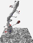
Fig. 1.4.1.46. 3D reconstruction of a spiny branchlet on EM background
Fig. 1.4.1.47. Spine apparatus in a dendritic spine neck
Fig. 1.4.1.48. Dense plate radiates from spine apparatus into punctum adhaerens
Fig. 1.4.1.49. 3D reconstruction of a dendritic spine of a Purkinje cell
Fig. 1.4.1.50. 3D reconstruction of mushroom spines with perforated synapses
Fig. 1.4.1.51. 3D reconstruction of a multisynaptic mushroom spine
Fig. 1.4.1.52. 3D reconstruction of an axon forming a synapse with a mushroom spine
Fig. 1.4.1.53. Apical dendrite of hippocampal pyramidal neuron
Fig. 1.4.1.54. Mushroom-shaped dendritic spines with perforated synapses
Fig. 1.4.1.55. Mushroom-shaped spine with spine apparatus
Fig. 1.4.1.56. 3D reconstructions of somatic spines
Fig. 1.4.1.57. 3D reconstructions of apical dendrite from hippocampal pyramidal neuron
Fig. 1.4.1.58. EM image indicating importance of 3D analysis
Fig. 1.4.1.59. 3D reconstruction of mushroom-shaped dendritic spine forming spinule
Fig. 1.4.1.60. 3D reconstruction of mushroom-shaped dendritic spine and astrocyte