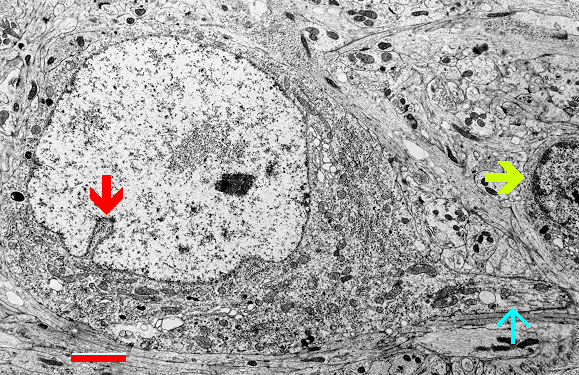
Fig. 1.2.09. The nucleus of the Golgi cell with indentation (red arrow). An axon initial segment is apparent at the cell base (blue arrow). Granule cell - yellow arrow. Scale = 3 µm. (Rabbit, cerebellar cortex). Download the high resolution image.
Fig. 1.2.09. The nucleus of the Golgi cell with indentation (red arrow). An axon initial segment is apparent at the cell base (blue arrow). Granule cell - yellow arrow. Scale = 3 µm. (Rabbit, cerebellar cortex). Download the high resolution image.