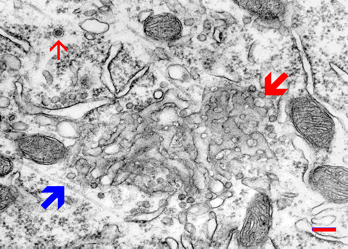
Fig. 1.1.3.02. Golgi apparatus in Purkinje neuron as it appears tangentionally viewed (red arrow). Neurosecretory granule (thin red arrow), microtubule (blue arrow). Scale = 200 nm. (Mouse, cerebellar cortex). Download the high resolution image.
Fig. 1.1.3.02. Golgi apparatus in Purkinje neuron as it appears tangentionally viewed (red arrow). Neurosecretory granule (thin red arrow), microtubule (blue arrow). Scale = 200 nm. (Mouse, cerebellar cortex). Download the high resolution image.