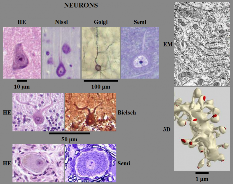
Fig. 0.1. From Light Microscopy to Ultrastructure and Three-Dimensional Reconstructions: Pyramidal cells of neocortex are shown in first line, Purkinje cells of cerebellar cortex in second line, ganglion cells of trigeminal ganglion in third line. Standard hematoxyline-eosin staining (HE) is compared with cresyl violet staining (Nissl), silver impregnations (Golgi, Bielschowski) and toluidine blue stained epoxy resin semithin sections (Semi). Part of pyramidal cell perikaryon is shown on electron micrograph (EM) and appearance of dendrite of Purkinje cell is shown on three-dimensional reconstruction (3D).
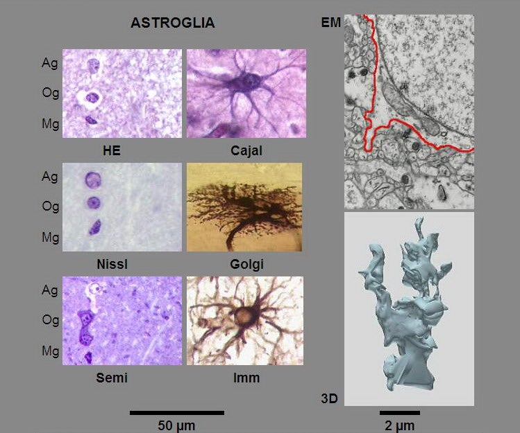
Fig. 0.2. From Light Microscopy to Ultrastructure and Three-Dimensional Reconstructions: Shapes of neuroglial nuclei (astrocyte - Ag, oligodendrocyte - Og, microgliocyte - Mg) are compared in standard hematoxyline-eosin staining (HE), cresyl violet staining (Nissl) and toluidine blue stained epoxy resin semithin section (Semi). Compare apearances of astrocytes in gold and silver impregnations (Cajal, Golgi) and immunohistochemical staining (with vimentin antibodies). Part of astrocyte cell body is shown on electron micrograph (EM) and appearance of astrocytic processes is shown on three-dimensional reconstruction (3D).
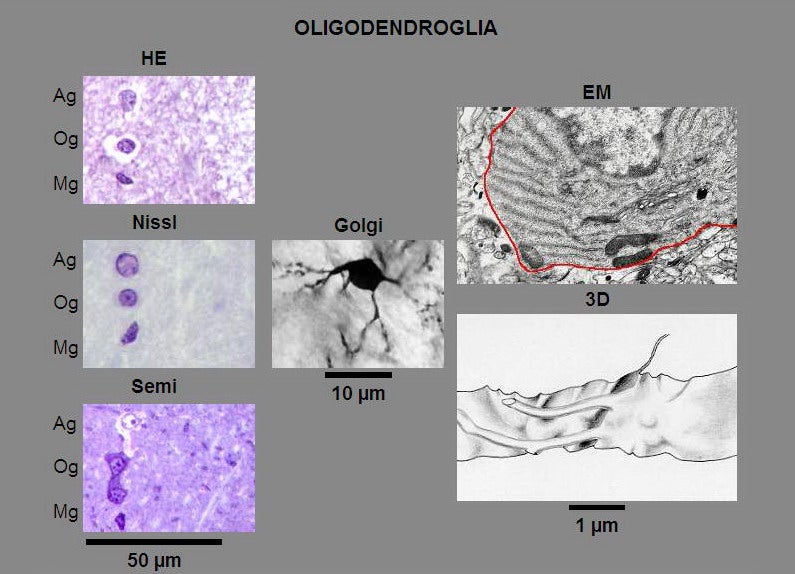
Fig. 0.3. From Light Microscopy to Ultrastructure and Three-Dimensional Reconstructions: Shapes of neuroglial nuclei (astrocyte - Ag, oligodendrocyte - Og, microgliocyte - Mg) are compared in standard hematoxyline-eosin staining (HE), cresyl violet staining (Nissl) and toluidine blue stained epoxy resin semithin section (Semi). Oligodendrocyte with its sparse processes is shown in silver impregnation (Golgi). Part of oligodendrocyte cell body is shown on electron micrograph (EM) and appearance of its processes is shown on three-dimensional reconstruction (3D).

Fig. 0.4. From Light Microscopy to Ultrastructure and Three-Dimensional Reconstructions: Shapes of neuroglial nuclei (astrocyte - Ag, oligodendrocyte - Og, microgliocyte - Mg) are compared in standard hematoxyline-eosin staining (HE), cresyl violet staining (Nissl) and toluidine blue stained epoxy resin semithin section (Semi). Microgliocyte with its processes is shown in silver impregnation (Hortega). Part of microgliocyte cell body is shown on electron micrograph (EM) and appearance of its process is shown on three-dimensional reconstruction (3D).
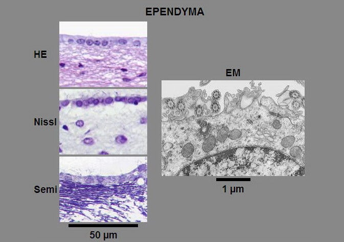
Fig. 0.5. From Light Microscopy to Ultrastructure: Appearances of ependyma cells are compared in standard hematoxyline-eosin staining (HE), cresyl violet staining (Nissl), toluidine blue stained epoxy resin semithin section (Semi) and electron micrograph (EM). Note kinocilia visible in Nissl and toluidine blue staining.
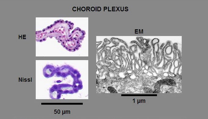
Fig. 0.6. From Light Microscopy to Ultrastructure: Appearance of choroid plexus is compared in standard hematoxyline-eosin staining (HE), cresyl violet staining (Nissl) and electron micrograph (EM).
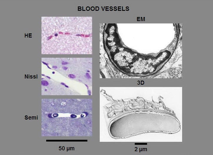
Fig. 0.7. From Light Microscopy to Ultrastructure and Three-Dimensional Reconstructions: Blood capillaries of neocortex are compared in standard hematoxyline-eosin staining (HE), cresyl violet staining (Nissl), toluidine blue stained epoxy resin semithin section (Semi) and electron micrograph (EM). Astrocyte process associated with capillary wall is visible on three-dimensional reconstruction (3D).
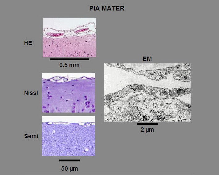
Fig. 0.8. From Light Microscopy to Ultrastructure: Appearance of pia mater of neocortex is compared in standard hematoxyline-eosin staining (HE), cresyl violet staining (Nissl), toluidine blue stained epoxy resin semithin section (Semi) and electron micrograph (EM). Note blood vessels in pia mater.
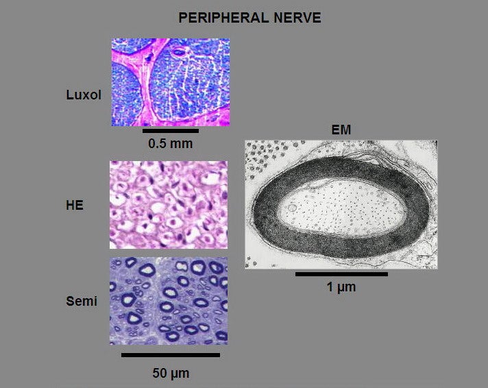
Fig. 0.9. From Light Microscopy to Ultrastructure: Appearances of myelinated axons in peripheral nerves are compared in standard hematoxyline-eosin staining (HE), Luxol fast blue staining (Luxol), toluidine blue stained epoxy resin semithin section (Semi) and electron micrograph (EM).